Category Archives: New Papers

piRNAs in the brain: epigenetics and memory

An exciting new paper in Cell, links Piwi-interacting RNAs (piRNAs) to long-term memory via the epigenetic regulation of gene expression by DNA methylation.

Two different novel findings are especially important: piRNAs had been thought to be a germline specific mode of genetic control, specifically a type of genetic immunity against the mobilisation of transposable elements. Rajasethupathy et al demonstrate that in the sea hare, Aplysia, piRNAs are expressed in the CNS and other somatic tissues. Secondly, this paper demonstrates specific dynamic de novo methylation of the promoter of a gene regulating neuronal plasticity in response to neurotransmitter- mediated excitation. This provides an epigenetic mechanism by which memories can persist by molecular encoding.

To investigate microRNAs expressed in the Aplysia CNS, Rajasethupathy et al had constructed a small RNA library. Surprisingly, they found that ~20% of the sequence reads from this library were ~28nt long, and showed a bias towards having 5′ uridine residue. This fitted with them being Piwi-interacting RNAs (piRNAs) rather than microRNAs. When they were mapped to the Aplysia genome, it was clear that the piRNAs were generated from piRNA clusters (see previous introductory post). After constructing more libraries and deep sequencing, Rajasethupathy et al. identified 372 distinct Aplysia piRNA clusters. Certain piRNAs are found far more commonly than surrounding piRNAs from the same cluster, indicating that an amplification process is occurring during piRNA biogenesis. Although overall piRNA content was highest in the ovotestes (ie the germline and associated somatic tissues), various piRNAs were found to be enriched in the CNS, as well as other somatic tissues analysed.

Consistent with the presence of piRNAs in the CNS, Rajasethupathy et al. were able to clone a full-length cDNA for Piwi protein from Aplysia CNS. Using an antibody against Piwi, they were then able to co-immunoprecipitate piRNAs with Piwi protein from the CNS. By separating cell nuclei from cytoplasm and then western and northern blotting against Piwi and piRNAs respectively, they showed that both were primarily found in nuclei.

To briefly comment on the piRNA aspect of this paper; a number of outstanding questions arise. In the best characterised model systems, piRNA amplification occurs by the ‘ping-pong’ mechanism in which reciprocal recognition and cleavage reactions between sense and antisense piRNAs complexed with the (cytoplasmic) Piwi-related proteins Aubergine and AGO3 (Drosophila terminology), leads to selective amplification of piRNAs with a tell-tale 10nt offset. Neither of these proteins, nor the 10nt offset, nor the ratio between sense and antisense piRNAs are mentioned by Rajasethupathy et al. I imagine that these questions would’ve been looked into and mentioned if found, therefore it seems that the mechanism of piRNA amplification in the Aplysia CNS is potentially novel.

To explore possible functions of piRNAs in the Aplysia CNS, the researchers used a co-culture system that monitors changes in synaptic plasticity in response to stimulation by the neurotransmitter serotonin (5HT). In this assay, two sensory neurons synapse with a motor neuron. Changes in the strength of one sensory-motor synapse are monitored by electrophysiological recording from the motor neuron. This system measures long-term facilitation (LTF): ie. changes in synaptic strength in response to 5HT stimulation. LTF is considered to be a memory-related phenomenon, however, it is contentious just how well it serves as a paradigm for long-term memory.

Knockdown of Piwi (and hence of complexed piRNAs), by the injection of antisense oligonucleotides into one of the sensory neurons, significantly impaired LTF, whilst overexpression of Piwi enhanced it. To investigate how these Piwi effects on LTF were mediated, Rajasethupathy et al. looked at the expression of proteins known to be regulating synaptic plasticity in response to Piwi knockdown. Only one of the assayed proteins was responsive: CREB2, a transcriptional repressor, known to be a major inhibitory constraint on LTF, was upregulated in response to Piwi knockdown. Interestingly, an even greater increase in CREB2 mRNA was observed.

The fact that Piwi knockdown led to an increase in CREB2 mRNA, and it’s nuclear localisation, suggested that rather than acting post-transcriptionally (ie by degrading mRNAs as in Drosophila), Piwi/piRNA complexes appeared to be inhibiting CREB2 gene expression at the DNA level.  It is known that in mice Piwi/piRNA complexes act to silence transcription by facilitating DNA methylation. Rajasethupathy et al therefore asked whether CREB2 regulation by Piwi occurred via DNA methylation.

The enzyme responsible for methylation of cytosine residues in CG dinucleotides, DNA methyltransferase (DNMT), was known to be expressed in the Aplysia CNS. Inhibition of DNMT activity (using a pharmacological reagent) led to a strong increase in the level of CREB2. In the normal LTF experiments, CREB2 levels are reduced 12 hours after exposure to 5HT and remain low until 48hrs. This downregulation of CREB2 was abolished when DNMT activity was inhibited, as was 5HT-dependent LTF. This led the researchers to search for CpG islands in the promoter region of CREB2 that could be sites for DNA methylation mediated transcriptional control. Indeed, they identified a CpG island in the CREB2 promoter region that normally exists in both methylated and unmethylated states. After 5HT exposure, this CpG island is almost entirely methylated, whilst in the presence of the DNMT inhibitor it becomes almost entirely unmethylated. This 5HT-dependent methylation of the CREB2 CpG island requires Piwi, as it was abolished when Piwi was inhibited. The authors then went on to search for candidate piRNAs that could be responsible for mediating this effect, by searching for those with complementarity to the CREB2 promoter. They identified 4, one of which, aca-piR-F, when knocked down caused an increse in CREB2 expression. Notably, Rajasethupathy et al. did not demonstrate the expected result that aca-piR-F knockdown would lead to demethylation of the CREB2 CpG island, although this experiment was surely attempted.

In conclusion, this paper offers a broad outline for a mechanism of memory encoding; it connects neurotransmitter synaptic stimulation with the stable epigenetic marking of the transcription state of an important regulator of neuronal plasticity, via the action of Piwi/piRNA complexes. It should be noted that ‘epigenetic’ is used in this context in a loose definition with reference only to a stable marking of cellular state. Strictly ‘epigenetic’ should refer to heritable non-genetic changes, but as neurons do not divide that is inapplicable. In this case DNA methylation is a relatively long-lasting mark. However, for instance the change in CREB2 expression with respect to 5HT-stimulated long term facilitation only lasts a couple of days – does this correspond to the level of methylation in the promoter? In which case, one gets an impression that DNA methylation and demethylation are highly dynamic processes in the Aplysia CNS. Currently there are three modes proposed to resolve the difficult question of how memories can persist for a long time, whilst the cellular components that must mediate them have a high rate of turnover: Prion-like synaptic marks, autoregulatory loops that can maintain a cell state whilst their components come and go, and epigenetic mechanisms that can alter gene expression in a long term manner. This paper shows a clear example of the latter mode, but the apparent dynamism of DNA methylation in this system suggests a lack of permanence.

Although I like the way this paper has ranged over a large terrain and connected so many disparate elements, by necessity it raises many questions and leaves many aspects of the work unmentioned. I’ve already mentioned some questions about Aplysia piRNAs; no doubt a fully annotated Aplysia genome will answer some of them. A few other questions and future directions spring to mind: The authors haven’t quite shown that DNA methylation is responsible for the transcriptional silencing at the CREB2 promoter, only correlated it. Likewise the mode by which Piwi/piRNA complexes act to promote DNA methylation is unclear. A wider question is the nature of DNA methylation in Aplysia and other invertebrates. Some invertebrates show virtually no DNA methylation (C. elegans, Drosophila) whilst the majority display mosaic patterns quite different from those found in vertebrates. This suggests functional differences, and without deeper knowledge of the role of DNA methylation in Aplysia it is difficult to guess how widely applicable these findings are in other systems. Likewise the finding that piRNAs are acting at the level of DNA methylation, previously only found in mammals, raises questions about the state of affairs in other invertebrate model systems. Also, do Aplysia piRNAs only act on DNA methylation, or post-transcriptionally aswell.? Future studies will no doubt also look at how this type of regulation corresponds to histone marks, and try to synthesise the different levels of regulation. Perhaps the most important take home message is that piRNAs are more than a germline specific immunity against tranposons. Just how widespread these other roles are is an open question.

Rajasethupathy, P., Antonov, I., Sheridan, R., Frey, S., Sander, C., Tuschl, T., & Kandel, E. (2012). A Role for Neuronal piRNAs in the Epigenetic Control of Memory-Related Synaptic Plasticity Cell, 149 (3), 693-707 DOI: 10.1016/j.cell.2012.02.057

sciseekclaimtoken-4fad81d2e9619

On Ribosomal Pausing

A new paper in Nature, describes how Shine-Dalgarno-like features in protein coding sequences, leads to bacterial ribosomes pausing during translation. Selection against ribosomal pausing leads to biases in codon usage and coding sequence evolution. Translational pausing represents a new level of regulatory control of gene expression.

Translation, the process by which the nucleotide sequence of mRNA transcripts is decoded and converted into amino acid sequence during protein synthesis, is carried out by ribosomes. Within the ribosome, transfer RNA molecules recognise specific trinucleotide codons on the mRNA, and add their cognate amino acids to nascent protein chains. In bacteria and archaea, ribosomes often recognise the translation start site with the help of a sequence 8 to 12 nucleotides upstream of it – the Shine-Dalgarno sequence (SD). It’s been known since the 1980s that different mRNAs are translated at different rates. The main reason for these differences was thought to be the concentration of rarer varieties of tRNA limiting the rate at which some transcripts could be decoded.

Li et al. have used a new technique, ribosome profiling, that maps ribosome occupancy along mRNAs. This has yielded high-resolution views of local translation rates on the entire protein coding transcriptome of E. coli and Bacillus subtilis.  Briefly put, mRNA fragments that have been protected from nuclease digestion by ribosomal binding, are ‘deep sequenced’. The concentration of these ribosome footprints equates to the ribosome occupancy throughout the transcriptome. The local translation rate is inversely related to ribosome occupancy.

Using this technique, Li et al. found many sites where ribosomal density is ten fold or more than the mean. They sought to correlate these translational pauses with specific codons. However, there was little link between occupancy of specifc codons and the abundance of their corresponding tRNAs. Therefore, the concentration of rare tRNAs is not responsible for much translational pausing under normal growth conditions.

To try to find sequence features that were linked to ribosomal pausing, the researchers then tried to correlate any trinucleotide sequences (independently of reading frame) with ribosome occupancy. They found that 6 different trinucleotide sequences, with features resembling Shine-Dalgarno sequences, did correlate with the position of paused ribosomes. This correlation was not found in the yeast, Saccharomyces cerevisiae; in agreement with eukaryotic ribosomes not using SD- anti-SD interactions.

Li et al. went on to show definitively that internal SD-like sequences are linked to translational pausing, by introducing a mutation into one such site and showing that ribosome occupancy was reduced. They also showed that peaks of ribosome occupancy, were caused by translational pausing, rather than attempted internal translational initiation.

As translational pausing limits the amount of free ribosomes, widespread internal SD-like sequences would limit the rate of protein synthesis, and hence the potential bacterial growth rate. In line with this, SD-like sequences in protein coding genes are disfavoured. Selection pressure against SD-like sequences is therefore a major factor in determining codon usage, and more especially the usage of codon pairs (SD sequences are 6/7 nt long).

Interestingly, the authors found that patterns of ribosome occupancy were conserved between orthologous genes in E. coli and B. subtilis. This reflects two different factors; firstly, coding is obviously constrained by protein’s functionality, but secondly it’s suggestive of translational pausing being exploited for functional purposes. Li et al. suggest a number of different ways in which ribosomal pausing can regulate gene expression. It’s known that internal SD-like sequences can promote regulated shifting of reading frame. Ribosome pausing may also modulate folding of nascent protein chains. Lastly, as transcription and translation are closely coupled in bacteria, ribosome occupancy may inhibit the formation of stem-loop structures that prevent transcriptional termination. It will be exciting to work out the extents to which these potential regulatory systems are active. Eukaryotic ribosomes do not use recognition of SD sequences, instead using the 5’ mRNA cap and the less well defined Kozak sequence for translational initiation. Does ribosome pausing occur in eukaryotes? and does it have functional significance?

Li, G., Oh, E., & Weissman, J. (2012). The anti-Shine–Dalgarno sequence drives translational pausing and codon choice in bacteria Nature, 484 (7395), 538-541 DOI: 10.1038/nature10965

microDNAs: small mammalian extrachromosomal circular DNAs

A new paper in Science, reports the detection of a new species of DNA in mammalian cells: microDNA. microDNAs are extrachromosomal circular DNA molecules, generally 200-400bp long, derived from non-repetitive genomic sequence. microDNAs appear to arise from microdeletions occurring in the 5’ ends of genes. This data implies widespread genetic variation with respect to microdeletions between somatic cells in mammals. 

To identify sites of intramolecular homologous recombination that could lead to genetic mosaicism in mammalian brains, Shibata et al. searched for the extrachromosomal circular DNAs (eccDNA) that could be produced. DNA was purified from embryonic mouse brains and linear DNA was degraded with a specific exonuclease. The remaining fraction was then amplified with an unbiased non-PCR technique (multiple displacement amplification). The linear products were then sheared into 500bp fragments, cloned, and sequenced. The majority of the clones in the library included repeated sequences, consistent with the products of rolling circle amplification of small circular DNAs. When these repeated sequences were searched against the mouse genome, they were only found once, showing that they were not produced by repetitive sequence (eg. transposable elements). To prove that these sequences were indeed derived from circular DNA molecules, PCR using outward directed primers designed from the repeated sequences was performed on both extrachromosomal and chromosomal DNA. If the template DNA was circular, PCR amplification should occur, if linear, it shouldn’t. This was (generally) the case, proving the existence of a population of extrachromosomal circular DNAs, a few hundred base pairs long, derived from unique portions of the chromosomal genome.

To further explore the nature and extent of this population of DNA molecules, Shibata et al. went on to purify eccDNA from a range of embryonic and adult mouse tissues, and from mouse and human cancer cell lines. After amplification and the sequencing of the ends of the generated fragments, they found that tens of thousands of unique genomic sequences yield extrachromosomal circular DNAs. The eccDNA from mouse tissues ranged from 80-2000bp in length, but most were between 200-400bp. Lengths of ~200bp and ~400bp were enriched in the mouse brain and liver populations. A similar pattern was detected in human cancer cell lines, but in these eccDNA populations an additional pattern of length distribution peaks at a 150bp periodicity was detected. As in the earlier experiment, the circular DNAs mapped to unique positions in the genome. To differentiate this population of eccDNA from previously reported longer forms derived from repetitive sequence, the authors termed them microDNAs.

Electron micrograph showing double-stranded (left) and single-stranded (right) microDNAs.

The researchers went on to directly visualise microDNA molecules by electron microscopy. Using a technique that specifically labels single stranded DNA, they discovered both double-stranded and single-stranded microDNAs were present in approximately equal measure.

Bioinformatic analysis of the sources of microDNAs revealed high enrichment for 5’ UTRs, exons, and CpG islands (regions upstream of genes where cytosine residues in CG dinucleotides are not methylated), suggesting that microDNAs are commonly derived from the 5’ ends of genes. microDNAs also have a higher percentage GC content than the average for the genome (55% as opposed to 45%). In a relatively high proportion of microDNAs, the researchers detected short direct repeats of 2-15bp of microhomology at the starts and ends of the molecules.

microDNAs could potentially be created by excision from chromosomal DNA, by replication of short stretches of DNA, or by reverse transcription of RNA molecules. Shibata et al. selected two genomic loci that yielded microDNAs and found that microdeletions do occur in these regions in some cells. The lengths and GC content of the microdeletions that they identified were in line with those found in microDNAs. The majority of the microdeletions displayed short stretches of microhomology at their excised ends.

These short direct repeats at the start and ends of microDNAs, and at their presumptive source microdeletions, suggest two possibilities for microDNA generation. Regions of microhomology could cause the DNA replication process to stall and switch template. Incorrect repair processes would then lead to the release of a microDNA. Alternatively, microhomology mediated repair processes could lead to the excision of a microDNA by intramolecular homologous recombination. The 150bp length periodicity detected in the cancer cell line microDNAs is suggestive of a link to nucleosomes (in which ~150bp of chromosomal DNA are wrapped around the histone core). A link to the position of nucleosomes (either in tightly bound nucleosomes causing replication problems or in facilitating microDNA circularisation) may explain the enrichment of microDNAs from the 5’ends of genes. Another suggestion made to explain the origin of ss microDNA, is that they could be formed by displaced Okazaki fragments (the short sections of replicated DNA formed on the lagging strand). All of these ideas are ‘hand wavey stuff’ but exciting avenues for future experiments nonetheless. A couple of obvious counter-arguments to these suggestions would be that microhomologies were only detected in 37% of microDNAs, and that the 150bp periodicity was only found in the cancer cell line microDNAs. A combination of the above putative modes of microDNA generation could be taking place, and microDNAs may be a heterogeneous population of molecules (as the presence of ss and ds DNAs suggests).

Perhaps the most striking conclusion of this paper is that the widespread generation of microDNAs by microdeletions yields large amounts of genetic variation between somatic cells. This mosaicism may well lead to functional differences between cells. What are implications of this mosaicism? Do microDNAs have any specific functions? Or are they simply a product of defective replication/repair processes? Are microDNAs only found in mammalian cells? Or are they more widespread (the researchers didn’t observe any in yeast cells)? It will be exciting to see future research attempt to answer these questions.

Shibata, Y., Kumar, P., Layer, R., Willcox, S., Gagan, J., Griffith, J., & Dutta, A. (2012). Extrachromosomal MicroDNAs and Chromosomal Microdeletions in Normal Tissues Science, 336 (6077), 82-86 DOI: 10.1126/science.1213307

Double-strand break interacting RNAs (diRNAs)

A new role for small RNAs in the repair of DNA double-strand breaks has been reported in Cell. Wei et al. have found diRNAs, derived from the vicinity of DNA double-strand breaks, in both Arabidopsis thaliana and human cells.

DNA double strand breaks (DSBs) are a particularly deleterious form of DNA damage as they can cause chromosomal translocations and induce cell death. To maintain the genome’s integrity, eukaryotic cells employ two different mechanisms of DSB repair. Non-homologous end joining (NHEJ) is an efficient mechanism that rapidly repairs DSBs without requiring an homologous template. However, NHEJ often causes insertions or deletions at the break site. Homologous recombination (HR) is a less error prone mechanism in which a sister chromatid is used as a template for repair. A specialised form of HR, single-strand annealing (SSA) that doesn’t require a sister chromatid can take place at repetitive sequences.

Wei et al. used an assay system that monitors DSB repair by SSA in the model plant Arabidopsis thaliana. A genetic cross causes a single DSB in an inactive reporter gene containing a repeat. SSA mediated repair restores the activity of the reporter gene and allows a quantitative and visible readout of DSB repair events. For instance, when this assay system is introduced (by crossing) into a genetic background mutant for atr (encoding a PI3 kinase known to be involved in DSB response), the researchers observed a large reduction in repair efficiency.

The first clue that suggested that small RNAs may be involved in double strand break repair came when they crossed their DSB repair assay system into lines mutant for Dicer-like proteins (DCL). Dicer and DCLs are responsible for the biogenesis of small RNAs (miRNAs and siRNAs) from double-stranded RNAs. Mutations in three different dcl genes (especially dcl3) all diminished the efficiency of DSB repair. The researchers therefore tried to examine whether small RNAs were produced from sequences adjacent to the DSB site. By probing northern blots with sequence flanking the DSB site, Wei et al detected a population of small RNAs approximately 21nt in length that were only present when DSBs had been induced. Deep sequencing (direct sequencing of RNA populations) revealed that these DSB-induced small RNAs (diRNAs) were specifically produced from the sequences flanking the DSB (approximately 800bp in each direction) and derived from both the sense and antisense strands in equal measure. By using a similar assay that monitored DSB repair by HR, they showed that diRNAs were also produced in this system.

In plants, a well characterised small RNA system mediates heterochromatic silencing of repetitive sequences by DNA methylation. Wei et al. used this pathway as a model to dissect the diRNA system. In the heterochromatic-siRNA system, single stranded RNA transcripts generated by the DNA-dependent RNA polymerase IV (Pol IV)  are converted to dsRNAs by the action of the RNA-dependent RNA polymerase 2 (RDR2). The dsRNAs are then cleaved into hc-siRNAs by Dicer-like proteins. When complexed with the Argonaute protein AGO4, hc-siRNAs direct de novo DNA methylation. By using the DSB assay system in backgrounds mutant for these factors and deep sequencing, Wei et al.  found that diRNA production requires the activity of Pol IV, RDR2 and RDR6 and DCLs, and that this pathway is under the control of DSB responsive kinase ATR. However, diRNA-mediated DSB repair does not involve RNA-directed DNA methylation pathway effector components such as AGO4. Instead, a different Argonaute protein AGO2 was found to complex diRNAs. Both diRNA accumulation and DSB repair were compromised in ago2 mutants.

Wei et al. went on to enquire as to whether diRNAs are involved in DSB repair in animals as well as plants. Using a similar HR mediated DSB repair assay in a human cell line, they showed small RNAs are also produced close to DSBs. Interestingly, whereas in plants the diRNAs were produced from sequences immediately neighbouring the DSB, in human cells they originated from a broader vicinity around the break site and not immediately adjacent. When Dicer or Ago2 were depleted in human cells DSB repair was compromised.

This paper has demonstrated the existence of a new class of small RNAs and their involvement in yet another important biological process. However, the details of how diRNAs act in DSB repair are completely unknown as yet. The authors suggest that diRNAs may guide histone modifications around the DSB site that facilitate DNA repair. Alternatively, diRNA-AGO2 complexes may be directly target DSB repair complexes to break sites. The assay systems used in this study only tested DSB repair by HR and SSA. It would be interesting to know whether diRNAs are also involved in DSB repair by NHEJ.

Wei, W., Ba, Z., Gao, M., Wu, Y., Ma, Y., Amiard, S., White, C., Danielsen, J., Yang, Y., & Qi, Y. (2012). A Role for Small RNAs in DNA Double-Strand Break Repair Cell DOI: 10.1016/j.cell.2012.03.002

Transposons and plasmids combine for bacterial chromosomal transfer.

A mechanism that facilitates the horizontal transfer of large segments of chromosomal DNA has been discovered in natural isolates of the pathogenic bacterium Yersinia pseudotuberculosis.

Although horizontal gene transfer (HGT) is recognised as a major force in bacterial evolution, and many mobile genetic elements underlying genomic plasticity have been characterised, the mechanisms by which genetic exchange of chromosomal DNA occurs in natural populations have remained largely hypothetical.

In experiments analysing the mobility of a pathogenicity island (HPI) in Yersinia pseudotuberculosis, Lesic and Carniel (2005) discovered that HPI could be transferred between natural isolates in a process that occurred optimally at 4˚C. However, this process didn’t require integration/excision machinery encoded in the HPI, and also transferred adjacent sequences (>46kb). To test whether this process was specific to the HPI region, Lesic et al inserted various antibiotic resistance markers equidistantly within the Y. pseudotuberculosis chromosome. When this strain was co-incubated with a naïve recipient strain, transfer of these markers was observed in ‘transconjugants’. None of the transconjugants acquired more than one of the antibiotic resistance loci, showing that the transferred chromosomal fragments were less that 1.5Mb in size. The transfer mechanism was also capable of mediating the transmission of a non-mobilisable plasmid (pUC4K). Together these results showed the existence of a mechanism for generalised DNA transfer that functioned at low temperature (termed GDT4).

Lesic et al. found that the GDT4 performing strain contained a very high molecular weight plasmid (≥100Mb). When this plasmid was removed (‘cured’) from the strain, GDT4 was abolished. They therefore termed this plasmid pGDT4. pGDT4 was transferred during transconjugation experiments and was able to confer the ability to transfer chromosomal DNA.

Electron micrograph of aggregating Y. pseudotuberculosis. White arrows point to bridge-like structures.

Sequencing of pGDT4 showed that it contained genes involved in conjugation (the process by which a pilus acts as a conduit for transfer of DNA between cells). When part of this conjugative machinery was deleted GDT4 ability was lost. Interestingly though, pili could not be observed by electron microscopy, and strong shaking of cultures (predicted to disrupt pilus-mediated interactions) had no effect. Instead, the bacteria were seen to tightly aggregate, and seemed to be connected by ‘bridges’.

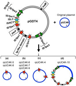
Organisation of pUC4K plasmids from transconjugants, showing novel IS insertions and IS mediated novel organisations.

pGDT4 also contains a number of Insertion Sequences (IS, short bacterial transposable elements). None of these IS were present on the bacterial chromosome or pUC4K (the non-mobilisable plasmid). After GDT4 transfer of pUC4K, it was observed the size of pUC4K was often altered. Of 10 transconjugant pUC4Ks measured, 3 were the original size, but (pGDT4 derived) IS insertions had lengthened the others. In 5 cases a single copy of ISYps1 had been acquired by pUC4K, whilst in 1 case a large section of pGDT4 had been inserted between two ISYps3 elements.

ISYps1 and ISYps3 (members of the IS6 and Tn3 families respectively) transpose by a mechanism termed replicative transposition. An intermediate stage during this mode of transposition is the formation of a ‘co-integrate’ in which the donor and target replicons (in this case pGDT4 and pUC4K) are fused by 2 copies of the transposon. The cointegrate is then resolved by homologous recombination, leaving a new copy of the transposon on the target and the original still in place on the donor.

Model describing ISYps1 transposition mediated mobilisation of pUC4K via cointegrate formation.

These observations suggested that GDT4 occurs by cointegrates of pGDT4 and other replicons (bacterial chromosome or other plasmids) being formed during IS transposition and then being transferred to recipient cells by conjugation. In further experiments, Lesic et al. confirmed that replicative transposition of ISYps1 was capable of  driving cointegrate formation between different plasmids in E.coli, that could also be transferred by conjugation.

This mode of chromosomal transfer is very similar to the ‘Hfr‘ system characterised in E. coli. In Hfr strains, transfer of chromosomal DNA occurs by conjugation because the conjugative plasmid ‘F’ becomes integrated into the chromosome. Integration of F occurs by homologous recombination at IS sequences shared between the plasmid and the bacterial chromosome. The difference in GDT4 is that no sequence identity between the two replicons is necessary. ISYps1 transposition does not seem to target any particular sequence. This makes this mode of chromosomal conjugative transfer less constrained than Hfr and potentially more powerful. It is notable that plasmids often carry a large number of IS. For instance, a plasmid in Shigella carries 93 copies of IS of 21 different types. The ability to confer chromosomal DNA transfer may be a selective advantage underlying plasmid/IS symbioses.

GDT4 was optimal at low temperature and low iron concentration. However, pGDT4 did not encode any known thermoregulators. This suggests that temperature sensitive pGDT4 conjugation is under the control host genes. GDT4 could therefore be a natural response to challenging growth conditions.

Lesic, B., Zouine, M., Ducos-Galand, M., Huon, C., Rosso, M., Prévost, M., Mazel, D., & Carniel, E. (2012). A Natural System of Chromosome Transfer in Yersinia pseudotuberculosis PLoS Genetics, 8 (3) DOI: 10.1371/journal.pgen.1002529

The Toxic Nano-Syringe of Vibrio cholerae.

Type VI secretion systems are used by bacteria to inject toxins into both bacterial competitors and host cells during pathogenesis. A new paper in Nature visualises these nanomachines and shows that they act by the swift contraction of a bacteriophage tail-like tube.

Bacterial type VI secretion systems (T6SS) are used to kill both eukaryotic and prokaryotic cells through the translocation of toxic proteins in a cell-cell contact-dependent process. T6SS encoding clusters of 15-20 genes are found in pathogenicity islands in the genomes of about a quarter of all sequenced gram –ve bacteria. Many T6SS proteins show sequence or structural homology with components of contractile phage tails. For instance, VgrG and Hcp, proteins secreted by T6SS, are structural homologues of a phage needle protein and phage tail tube protein respectively. Likewise, two Vibrio cholerae T6SS components VipA and VipB form tubular structures that resemble the tail sheath of T4 bacteriophage.

To investigate the role of VipA in T6SS, Basler et al. tagged VipA with a green fluorescent protein (GFP) and visualised it’s dynamics in cells. They found that the VipA-GFP fusion proteins were associated with long straight structures in the cell. These were often as long as the width of the cell (0.75-1µm) and varied in number between 0 and 5. By using time-lapse imaging, the researchers found that these putative sheath structures underwent a dynamic pattern of assembly, contraction and disassembly. The extended sheath assembled at a rate of 20-30 s µm-1. Sheaths then contracted to about 50% of their extended length in approximately 5ms. The contracted sheath was then disassembled in 30-60s.

Electron cryotomographic slices of V. cholerae showing Type VI secretion systems in the cytosol. In the left panel the T6SS is in the extended form, in the right it is contracted.


Basler et al. went on to visualise the T6SS sheaths directly using electron cryotomography. This discerned long straight tubular structures that existed in two conformations: A longer and thinner extended structure and a shorter and wider contracted form (see figure). The tubes were connected to the inner membrane by a flared bell-shaped base. Distal to the base in the extended T6SS structures was ‘conical-shaped density’ that crossed the periplasm and protruded through the outer membrane.

The results described in this paper are consistent with a model of T6SS action in which Hcp forms an inner tube within the VipA/VipB sheath. The Hcp tube, tipped with a VgrG ‘needle’ is fired into the target cell membrane by contraction of the T6SS sheath. Thus the energy captured by conformational change of the VipA/VipB polymeric sheath transports the toxic proteins through the cell membrane.

Basler, M., Pilhofer, M., Henderson, G., Jensen, G., & Mekalanos, J. (2012). Type VI secretion requires a dynamic contractile phage tail-like structure Nature, 483 (7388), 182-186 DOI: 10.1038/nature10846

Amyloid-like oligomers and long-term memory.

A new paper in Cell, shows that the formation of amyloid-like oligomers of a RNA-binding protein in synaptic membranes is necessary for the persistence of memories in Drosophila.

If memory is the maintenance of learning associated changes in synaptic efficacy and number, and these synaptic modifications are associated with changes in protein composition, how do they persist for years when individual protein molecules are turned over in days?

Cytoplasmic polyadenylation element-binding proteins (CPEBs) are a family of RNA-binding proteins that have been shown to regulate activity-dependent protein synthesis at synapses. A Drosophila CPEB, Orb2, has been shown to be required for the long-term persistence of memory (Keleman et al.). Previous studies on a CPEB from the sea slug Aplysia, had shown that it had prion-like properties, in that it could exist in two conformational states: a monomeric form and a self-perpetuating multimeric form with amyloid-like properties. Synaptic stimulation leads to the conversion of Aplysia CPEB into the multimeric form, and the formation of a self-sustaining synaptic mark. Blocking the activity of multimeric ApCPEB inhibited the long-term facilitation of a synaptic response (Si et al.).  Based on these Aplysia studies, Majumdar et al. hypothesised that Orb2’s role in Drosophila long-term memory could also be mediated by activity-dependent formation of amyloid-like oligomers.

Purifying Orb2 from Drosophila head extracts, Majumdar et al. show that it either exists in a monomeric form, or in oligomers consisting of between two and six Orb2 molecules. Orb2 oligomers are resistant to denaturation by heat or detergents, showing that they have amyloid-like characteristics. The oligomeric form of Orb2 appears to be localised to synaptic membranes, whilst the monomeric form is mainly found in cell bodies. Stimulation of neurons involved in memory formation increased the level of oligomeric Orb2.

Majumdar et al. go on to show that there are two different isoforms of Orb2 involved in the formation of Orb2 oligomers, Orb2A and Orb2B. Although the Orb2 oligomers found in vivo were mainly composed of Orb2B, the researchers found Orb2 oligomerisation requires Orb2A. Both isoforms contain a glutamine rich unstructured prion-like domain that acts as a substrate for oligomerisation, but a short 8 amino acid N-terminal domain of Orb2A catalyses the oligomerisation reaction. Orb2A appears to regulate the formation of amyloid-like Orb2 oligomers.

The previous study (Keleman et al.) that showed that Orb2 was involved in long-term memory stabilisation had used a technique that reduced the Orb2 concentration. By generating flies that carried a single point mutation in the Orb2A N-terminal domain, specifically impairing Orb2 oligomerisation, Majumdar et al. went on to show Orb2 oligomerisation is necessary for long-term memory persistence.

This work is exciting on two different fronts. It shows a mechanism for the generation of stable synaptic marks by the formation of amyloid-like oligomers. This could be a more widespread phenomenon underlying memory formation and persistence. More broadly, this study yields insight into potential physiological roles for amyloid formation and prion-like mechanisms.

Insoluble amyloid-like aggregations are a common pathological characteristic in human neurodegenerative conditions such as Alzheimer’s disease and Parkinson’s disease. In the case of prion diseases such as Creutzfeld-Jacob disease, amyloidogenic forms of Prion-Protein propagate the disease by seeding the conformational changes to the non-pathogenic form. The authors of the paper do not call the Orb2 oligomers prions, as the mechanisms underlying oligomerisation are not understood. However, in the case of ApCPEB, multimerisation was shown to be self-sustaining, and hence prion-like.

Is there a link between the preponderance of amyloid associated neurodegenerative diseases and the role of amyloid-like oligomers in memory? It is probably unwise to extrapolate too far from these results. However, one could postulate that if this is a widespread mechanism of memory formation, amyloid associated diseases could be more likely to affect the brain. Likewise, if amyloid-like protein multimerisation has normal physiological roles in the brain, it could be a more permissive environment for amyloid formation than other tissues.

Majumdar, A., Cesario, W., White-Grindley, E., Jiang, H., Ren, F., Khan, M., Li, L., Choi, E., Kannan, K., Guo, F., Unruh, J., Slaughter, B., & Si, K. (2012). Critical Role of Amyloid-like Oligomers of Drosophila Orb2 in the Persistence of Memory Cell, 148 (3), 515-529 DOI: 10.1016/j.cell.2012.01.004

Si, K., Choi, Y., White-Grindley, E., Majumdar, A., & Kandel, E. (2010). Aplysia CPEB Can Form Prion-like Multimers in Sensory Neurons that Contribute to Long-Term Facilitation Cell, 140 (3), 421-435 DOI: 10.1016/j.cell.2010.01.008

Keleman, K., Krüttner, S., Alenius, M., & Dickson, B. (2007). Function of the Drosophila CPEB protein Orb2 in long-term courtship memory Nature Neuroscience, 10 (12), 1587-1593 DOI: 10.1038/nn1996

A novel gene transfer agent from Bartonella

A gene transfer agent (GTA) that preferentially packages host adaptability genes has been discovered in the pathogenic bacterial genus Bartonella.

Bartonella are vector borne, facultative intracellular bacteria that infect erythrocytes and endothelial cells in various mammalian species. Berglund et al. sequenced the genome of the rodent infecting species Bartonella grahamii, and compared it with various other sequenced Bartonella genomes. Rodent associated Bartonella species had a larger amount of imported genes than other species. Most of these horizontally transferred genes were either bacteriophage derived, or encoded proteins involved in secretion or transport systems. A large proportion of the imported genes were located in a 461kb segment of the genome that the authors termed the chromosomal high plasticity zone.

The chromosomal high plasticity zone was present in all Bartonella species, but was most expanded in the rodent associated species. It showed similarities to auxiliary replicons (ie large extra-chromosomal elements) from related species.  Genes encoding type IV and type V secretion systems, that mediate bacterial invasion of erythrocytes and endothelial cells, were ancestrally located in the chromosomal high plasticity zone.

Bacteriophage particles were observed in Bartonella cultures. When these phage were isolated it was found that they contained proteins encoded at a cluster of phage genes in the high plasticity zone (phage cluster II), and packaged 14kb strands of DNA. By hybridisation of the phage encapsulated DNA against microarrays covering the bacterial chromosome, it was found that the phage contained DNA from the entire bacterial genome but that DNA from the high plasticity zone was overrepresented. The peak of the hybridisation signal was located at another cluster of phage genes within the high plasticity zone (phage cluster III).

It appears that auxiliary DNA replication from a phage derived origin of replication within the high plasticity zone is amplifying the Bartonella genome. The products of this ‘run-off’ replication are then being packaged into phage-like gene transfer agents. This is the first time a coupling of run-off replication and GTAs has been demonstrated. Previously discovered GTAs have packaged chromosomal DNA randomly, but in this instance the packaged DNA is biased towards DNA from the high plasticity zone, which is enriched for host adaptability genes.

The genes of the Bartonella GTA didn’t show any similarity to the RcGTA-like agents that are found in many other alpha-proteobacteria. Interestingly, Bartonella GTA-like clusters were identified in some other Rhizobiales species that also possessed RcGTA-like clusters. It would be interesting to enquire as to whether the GTAs in these species are also associated with run-off replication.

The authors suggest that the linkage of run-off replication and the GTA is selectively advantageous, in that not only is the rapid spread of new genes facilitated, but so is gene diversification by recombination.

 Berglund, E., Frank, A., Calteau, A., Vinnere Pettersson, O., Granberg, F., Eriksson, A., Näslund, K., Holmberg, M., Lindroos, H., & Andersson, S. (2009). Run-Off Replication of Host-Adaptability Genes Is Associated with Gene Transfer Agents in the Genome of Mouse-Infecting Bartonella grahamii PLoS Genetics, 5 (7) DOI: 10.1371/journal.pgen.1000546

Linking a lincRNA to active chromatin

Wang et al show that a lincRNA encoded at one end of the HOXA gene cluster acts as a transcriptional enhancer, necessary for the translation of high order chromosomal structure into a transcriptionally active chromatin state.

Hox genes encode transcription factors that determine positional identities along the anterior-posterior (a-p) body axis and along the proximal-distal (p-d) axes of appendages. In vertebrates, Hox genes are found in four clusters, in which their 3′-5′ genomic arrangement mirrors their a-p and p-d expression patterns. For instance, genes found at 3′ end of the HOXA cluster such as HOXA1 and HOXA2 are necessary for specifying positional identities in hindbrain, whilst genes found at the 5′ end of the cluster such as HOXA13 and HOXA11 determine distal elements of the limbs.

To investigate how this genomic colinearity is translated into differential p-d expression patterns, Wang et al analysed chromosomal conformation at the HOXA locus in different human fibroblast cells. In distally derived cells (foreskin or foot fibroblasts) the 5′ end of the HOXA cluster displayed a compact and looped conformation, whilst the 3′ end seemed largely linear. An opposite conformation was found in proximally derived cells (lung fibroblasts). These findings correlated with the presence of specific histone post-translational modifications (PTMs). Areas of high chromatin interactions showed high levels of trimethylated histone H3 lysine 4 (H3K4me3) (associated with transcriptionally active chromatin), and low levels of histone H3 lysine 27 trimethylation (H3K27me3) (associated with transcriptionally silent chromatin).

histone lysine methylation states across the HOXA cluster compared between distal and proximal cells.

A lincRNA named HOTTIP is encoded at the 5′ end of the HOXA locus. Analysis of it’s expression revealed that it is transcribed in posterior and distal territories, in a manner similar to nearby HOXA genes. When HOTTIP RNA was depleted (using small interfering RNAs) in distal cells, expression of 5′ HOXA genes was abrogated. HOXA genes nearest the HOTTIP gene were the most effected, however transcriptional activity over 40kb of the HOXA locus was lessened. HOTTIP RNA depletion did not effect the expression of other genes tested, such as the highly homologous HOXD genes.

HOTTIP RNA depleted distal cells did not show any changes to the higher order chromosomal conformation at the HOXA locus, however they did display a broad loss of H3K4me3 at the 5′ end of the cluster. This was not mirrored by a concomitant gain of H3K27me3. Therefore it appears the loss of 5′ HOXA gene expression upon HOTTIP RNA depletion is linked to the loss of H3K4me3.

Effects of HOTTIP RNA depletion on histone lysine methylation states.

H3K4 methylation and it’s maintenance are mediated by protein complexes composed of lysine methyltransferases such as MLL1 and associated proteins like WDR5. Analysis of MLL1 and WDR5 occupancy in the HOXA cluster of distal fibroblasts revealed occupancy peaks at the transcriptional start sites of the 5′ located genes. These occupancy peaks disappeared when HOTTIP RNA was knocked down.Wang et al go on to show that HOTTIP RNA physically interacts with WDR5 protein.

Effects of HOTTIP RNA depletion on occupancy of MLL1 and WRD5.

A number of lines of evidence suggest that HOTTIP RNA acts in cis to regulate 5′ HOXA genes. For instance, HOTTIP RNA is expressed at very low copy number, and depletion did not have any effect on the HOXD locus. Retroviral insertion driven overexpression of HOTTIP RNA did not ectopically activate HOXA genes, nor could it rescue depletion of the endogenous HOTTIP RNA.

Tying these results together yields a model in which chromosomal looping brings HOTTIP RNA (specifically bound to the HOTTIP gene) into contact with the 5′ HOXA genes. HOTTIP lincRNA binds and targets WDR5/MLL complexes to the 5′ HOXA locus, creating a domain of H3K4me3 and transcriptional activation.

lincRNAs are probably a heterogeneous class of molecules, as they are arbitrarily defined on the basis of length. These results however, suggest what could be quite a common mechanism for how lincRNAs implicated in enhancer function can effect gene activation and program chromatin states.

See also: lincRNAs in development and evolution

Wang, K., Yang, Y., Liu, B., Sanyal, A., Corces-Zimmerman, R., Chen, Y., Lajoie, B., Protacio, A., Flynn, R., Gupta, R., Wysocka, J., Lei, M., Dekker, J., Helms, J., & Chang, H. (2011). A long noncoding RNA maintains active chromatin to coordinate homeotic gene expression Nature, 472 (7341), 120-124 DOI: 10.1038/nature09819

Lysine Crotonylation and the Histone Code

A recent study has identified 67 new histone modifications, bringing the current total of known histone marks to 163. Two new classes of modification were discovered: lysine crotonylation and tyrosine hydroxylation. Tan et al go on to show that crotonylated lysine marks active promoters and potentially plays an important role in male germ cell differentiation.

Eukaryotic chromosomal DNA is condensed by being wound around octamers of histone proteins to form nucleosomes. Post-translational modifications (PTMs) of histones can modulate chromatin structure, altering its biological activity (for example it’s transcription status). Different combinations of histone proteins and their PTMs are found through the genome and between different cell types. Deciphering this ‘histone code’ is crucial to our understanding of cellular regulation and differentiation, and is therefore the focus of huge amounts of current biological research.

Prior to this new paper at least twelve different types of histone PTM, at over sixty different amino acid residues had been reported. These include the most commonly discussed such as methylation and acetylation, as well as esoterica like citrullination. By performing a highly comprehensive survey of histone PTMs based on mass spectrometry, Tan et al have identified two new types of modification and 67 new histone marks.

The structure of the nucleosome. The four core histones are in different colours. Their N terminal tails are protruding from the nucleosome.

Nucleosomal cores consist of histone octamers containing two molecules each of histones H2A, H2B, H3, and H4. Interactions between histone proteins and between histones and DNA are generally mediated within the globular core domains of the histone proteins, whilst their N-terminal tails protrude from the nucleosome and have been considered the primary sites for post-translational modifications. However, this new study identified many histone PTMs within the globular cores, suggesting that previous methods of PTM identification have been biased against their discovery.

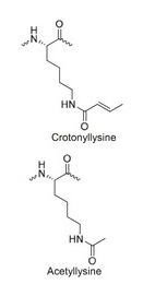
Tan et al also report further characterisation of one of the new types of histone PTM: lysine crotonylation (KCr). Crotonylation was found at 28 different lysine residues from all four core histones and the linker histone H1. KCr was detected in histones isolated from yeast, nematodes and fruit flies, as well as mice and humans.

Using an antibody that recognised all lysine crotonylation, chromatin immunoprecipitation followed by sequencing (ChIP-seq) showed that histone KCr was associated with active chromatin and was particularly enriched at promoter and enhancer regions.

Tan et al went on to find that during mouse spermatogenesis histone KCr is highly enriched in post-meiotic spermatids, coinciding with a general transcriptional shutdown. By using ChIP-seq in combination with transcriptomic data, they showed that KCr was marking a group of genes on the sex chromosomes that are transcriptionally active, whilst the rest of the sex chromosome is inactivated.

Lysine crotonylation appears to be an important new PTM adding even more complexity to an already complex field of study. The comprehensiveness of the technique employed for PTM identification used in this study, however, suggests that there may not be too many more histone marks to add to the list. The next questions to ask will be whether crotonylation of different lysine residues correlates with different biological events? What enzymes are responsible for the addition and removal of crotonyl modification? And what effects does the disruption of their activity have? What proteins interact with KCr? As can be ascertained from this taster, deciphering the histone code is going to keep a lot of people busy for a long time.

Tan, M., Luo, H., Lee, S., Jin, F., Yang, J., Montellier, E., Buchou, T., Cheng, Z., Rousseaux, S., Rajagopal, N., Lu, Z., Ye, Z., Zhu, Q., Wysocka, J., Ye, Y., Khochbin, S., Ren, B., & Zhao, Y. (2011). Identification of 67 Histone Marks and Histone Lysine Crotonylation as a New Type of Histone Modification Cell, 146 (6), 1016-1028 DOI: 10.1016/j.cell.2011.08.008